PET imaging
Imaging the state of boron accumulation in tumors and determining whether a given patient is a candidate for BNCT
Before treatment by BNCT, it is necessary to verify that boron (10B) has accumulated in the malignant tumor targeted for treatment but not in surrounding healthy tissue. The amount of 10B accumulation in the tumor directly affects the absorbed dose when irradiated with neutrons. Additionally, verifying low accumulation in healthy tissue enables BNCT to be administered safely with minimal adverse effects.
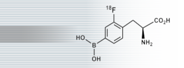
Positron emission tomography (PET) can be used to measure the extent to which a certain compound has accumulated in a patient’s internal organs. PET testing consists of administering a minuscule amount of a radioactive compound or pharmaceutical and then imaging the patient’s entire body. By marking the carrier that transports boron (10B) to the tumor with a radioactive nuclide and administering it to the patient, it is possible to quantitatively estimate the amount of boron in an individual patient’s tumor and surrounding tissue.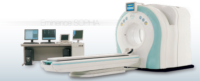
Borono-phenylalanine, a boron compound with a high propensity to accumulate in tumors
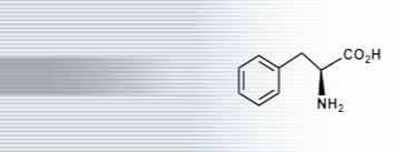
Phenylalanine, an aromatic amino acid, is metabolized more quickly in tumor cells that are growing rapidly. Since the compound cannot be synthesized inside cells, it is absorbed in large volumes from the blood.
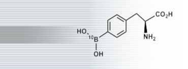
Borono-phenylalanine (BPA), derived by marking phenylalanine with 10B boron, is also taken up in large volumes by tumors. In BNCT, BPA is used as the principal boron carrier.
The compound 18F-fluoro-borono-phenylalanine (FBPA), derived by labeling BPA with the radioactive nuclide 18F, is used to image the distribution of BPA in the body using PET. Like BPA, FBPA also has a high propensity to accumulate in tumors.